Department is equipped with the latest X Ray machines: stationary digital machineDuoDiagnost, three mobile devices Practix 300 (located in intensive care unit for children and adults), spiral CT scanner Philips Brilliance CT64 with injector Stellant.
Digital X Ray research reduces radiation exposure to the patient and staff; X Ray study are giving a complete picture of the patient. The digital image can be recorded and transferred by network.
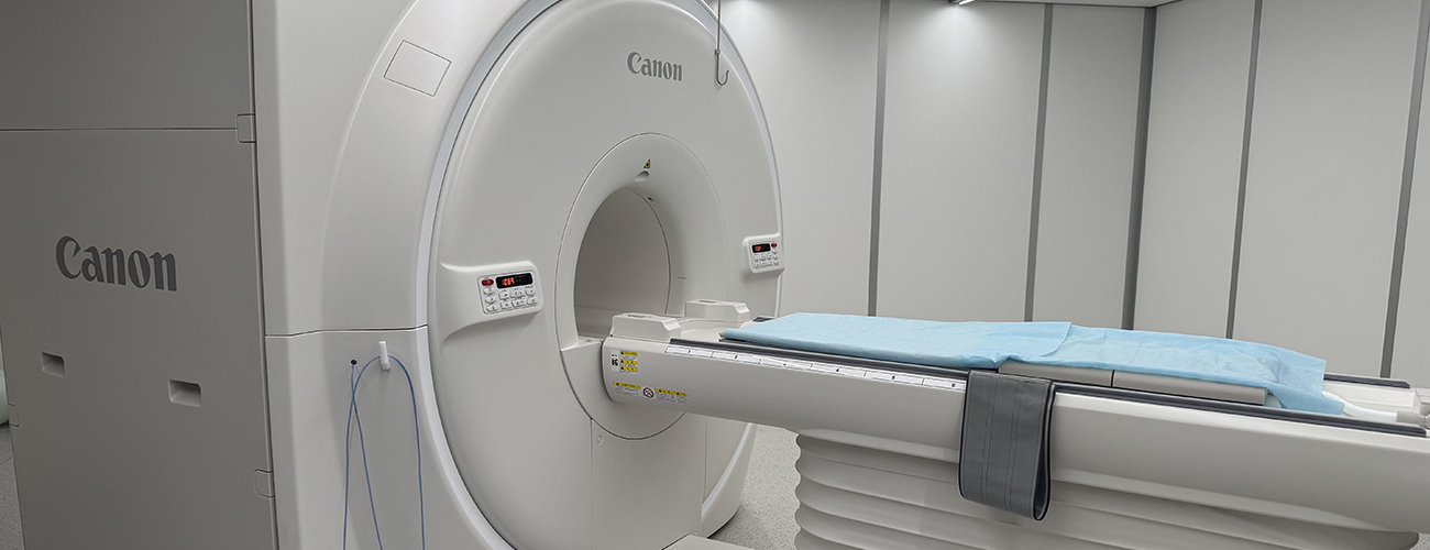
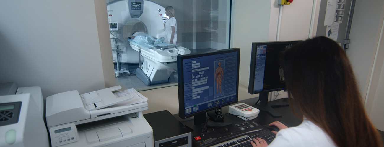
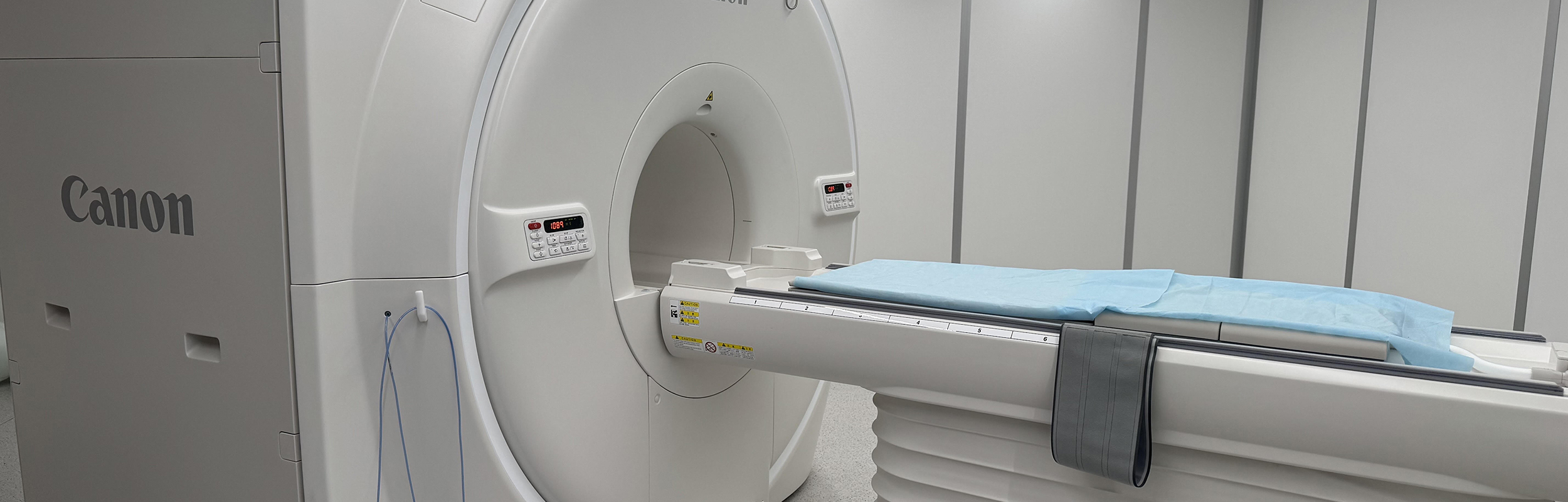
CT scanner Philips Brilliance CT 64 gives possibility to have comprehensive study of all the organs and systems for the 10s with slice thickness of 0.5 mm. Technical characteristics allow to perform the most sophisticated non-invasive angiographic study of any location. Part of the diagnostic deppartment is the latest generation graphics station from the company Philips, which named Brilliance Workstation. Brilliance Workstation allows us to study not only the layered cuts, but also to perform post-processing of the data with the performance of complex reconstructions which can schedule surgery. Specialized studies are SKT-coronary angiography, SKT-aortography, the study of brain vessels, research vessels of the lower extremities.
– MSCT coronary arteries, including control stent patency
– MSCT bypass angiography, aortography
– MSCT skull and brain without contrast
– MSCT of the brain and neck / with contrast
– MDCT of the chest
– MDCT angiography
– MDCT of the abdomen and retroperitoneum
– MDCT of the abdomen with a double-contrast
– MSCT spine
– MSCT of the pelvis and pelvic organs (LIB)
– MSCT joints of upper and lower extremities
– MDCT angiography of the lower limbs
MSCT angiography to diagnose different diseases
Studies using the contrast:
MSCT angiography: the aorta (thoracic and abdominal) and peripheral vascular disease (brain, neck, pulmonary arteries, upper and lower extremities) are conducted to diagnose the following diseases:
• Aorta: arch anomalies, coarctation, aneurysms, stratification, stenosis, occlusion, artery, traumatic ruptures, postoperative study (artificial valves, aortic)
• Carotid artery stenosis, aneurysms, bundle, twisted turn, looping
• Celiac artery stenosis syndrome, median arcuate ligament
• Hepatic artery: clarify the anatomy before surgery, detection of stenosis or occlusion after liver transplantation
• Mesenteric vessels: chronic, acute ischemia, aneurysms
• Renal artery stenosis, developmental abnormalities, fibromuscular dysplasia
• The arteries of the upper and lower extremities: occlusive disease, aneurysms, postural compression of the subclavian artery steal syndrome
• Postoperative complications: bleeding, infection, thrombosis, graft, anastomotic aneurysms